
Humans around the world see doctors everyday. We go to doctors for many reasons such as to have routine checkups, get medicine when we feel sick, get injuries repaired, or even surgery, but we are not the only ones. Animals that are different from us get hurt and sick too and humans have stepped up to take care of them. They are doctors just like ours but they treat other animals, not humans. These people are called veterinarians and we recently had to take one of our corn snakes, William Snakespeare, to see the wonderful veterinarians and surgeons at BloomingPaws and Avian and Exotic Animal Clinic of Indianapolis (AEACI).
William’s symptoms first manifested early September of 2017. She had been constipated and had a bulge growing near her vent (opening where all poop, pee and gametes are released) (see Image I). On September 4, William had regurgitated a partially digested mouse. Unfortunately, we do not have a photo of this, but we could tell it was a regurgitation rather than feces because 1) it was only partially digested. If it had gone through the entire digestive track, there would not be complete pieces of the mouse and 2) the regurgitation was covered in mucus, abnormal for feces, but normal for regurgitation and a sign that she can no longer push more food through her system. We immediately called our vet.
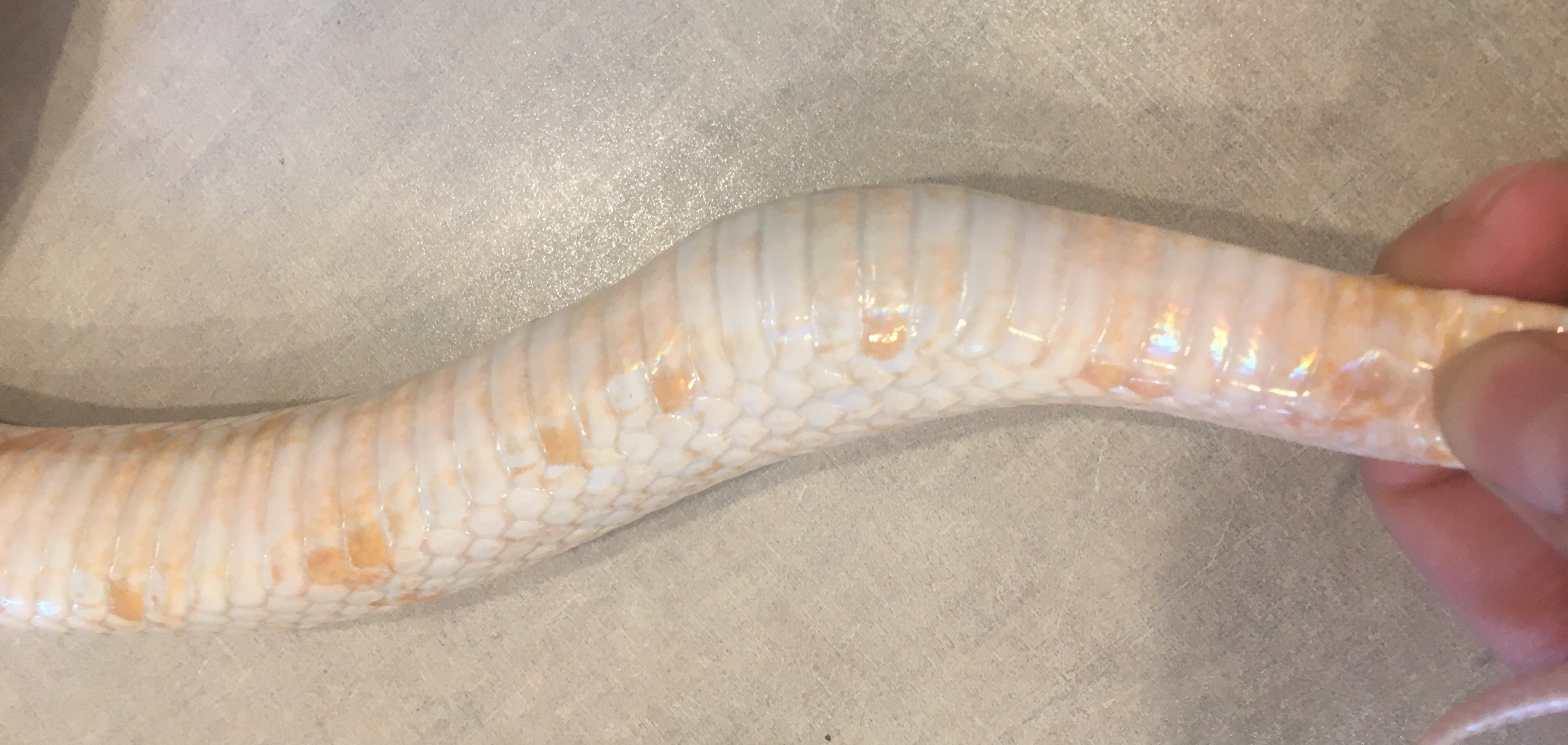
Image I: This is a lateral view (sideways) of the bulge that was seen by William’s vent. You can see the bulge right where her curve is. Taken by Sam Couch at WonderLab 9/5/17.
The first vet we went to was at BloomingPaws here in town. They took radiographs (x-rays) and what we saw was a bit scary (see Image II). What this shows is that the bulge was a mass that was keeping feces from passing through. You can also see that she has feces filling almost 16 cm of her gastrointestinal (GI) track You can tell what is what by the differences in color and texture of each part.
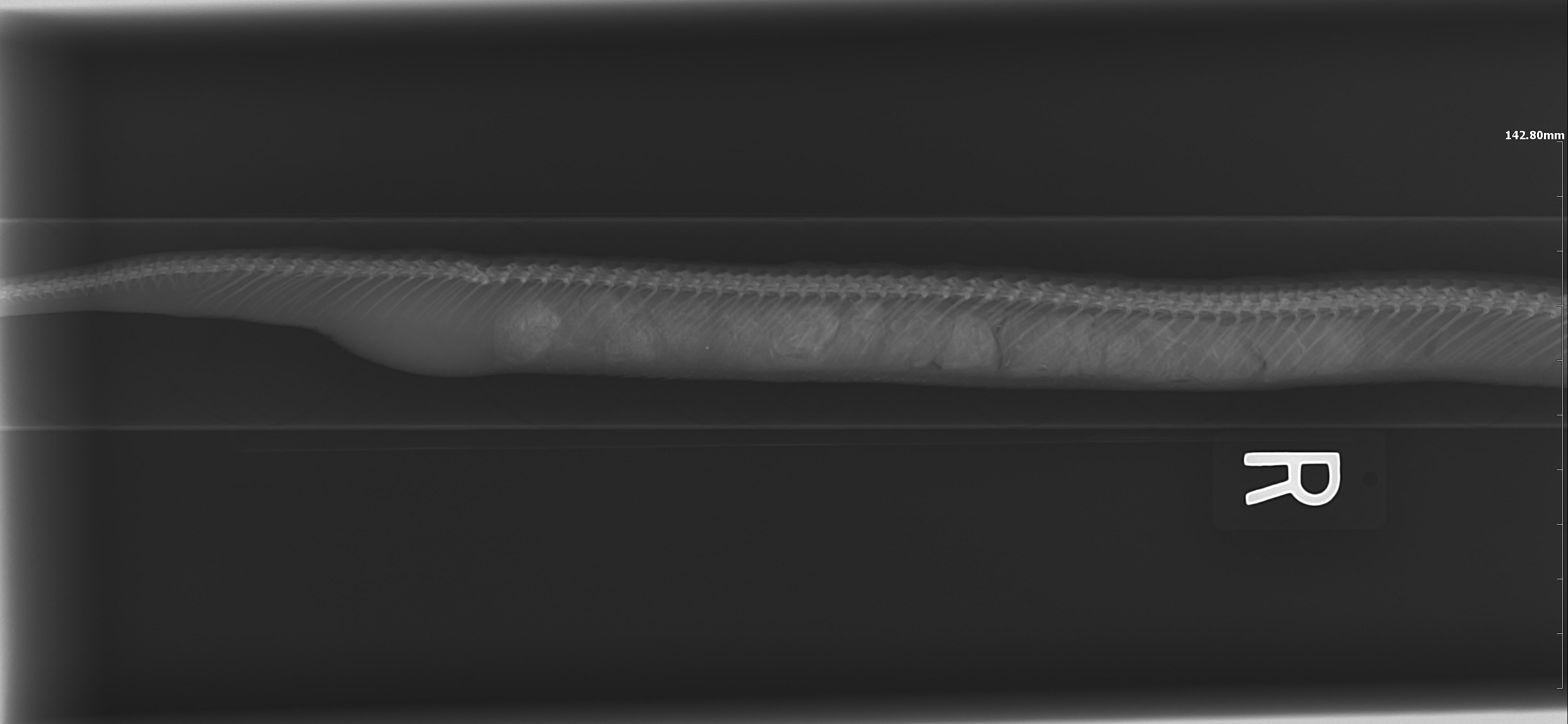
Image II: The mass is the medium colored curve on the left side of the image. The feces is the lightest, most textured portion that runs from the end of the mass through about 16 cm of GI track. Where the feces and mass meet. you can see a dip in her spine. We briefly discussed this as being the cause but the evidence for the mass, and the fact that there was no associated incident with the spine lead us to the hunch that the mass was the primary issue. Radiograph was taken at BloomingPaws 9/7/17.
After two weeks of massage and physical therapy with me and an unsuccessful enema later, William was taken into surgery at AEACI. It turns out she had a mass on her kidney that was blocking her vent from allowing the feces to pass. The great surgeons in Indy were able to remove the entire mass with little chance of recurrence. They were able to confidently get the mass because snakes like this are able to lose up to about 70% of their kidney before seeing any signs of renal failure which allowed the surgeons to get a very clean scrape. They say even if it does recur, the growth rate of something like this is so slow in ectotherms (animals that do not produce their own heat aka cold blooded) that we wouldn’t see symptoms in her lifetime!
We saw results almost immediately! The doctors said that her GI track was already contracting as they were closing her up after surgery. In fact, she had the largest bowel movement I have ever seen from corn snake on the drive home the next morning! It was comparable to that of a beagle sized dog which is significant considering snakes eat only once every two weeks and dogs eat multiple times a day! This puts the situation in a bit of perspective.
She came out of surgery with only three itty bitty stitches along her side and no signs of a mass (See image III). The sutures were not dissolvable and had to remain in for 6 weeks. Dissolvable sutures do not work on ectotherms because their metabolic rates are so low. These sutures are actually in the epidermis (top layer skin) that is underneath the scales. This means she will still be able to shed, which she did do once while she had stitches (See Image IV). This posed a bit of a problem because as you can see the stitches held in some of the old skin. We pulled what we could and let the doctors remove the rest on November 6 when she got all of her stitches removed (See Image V)!
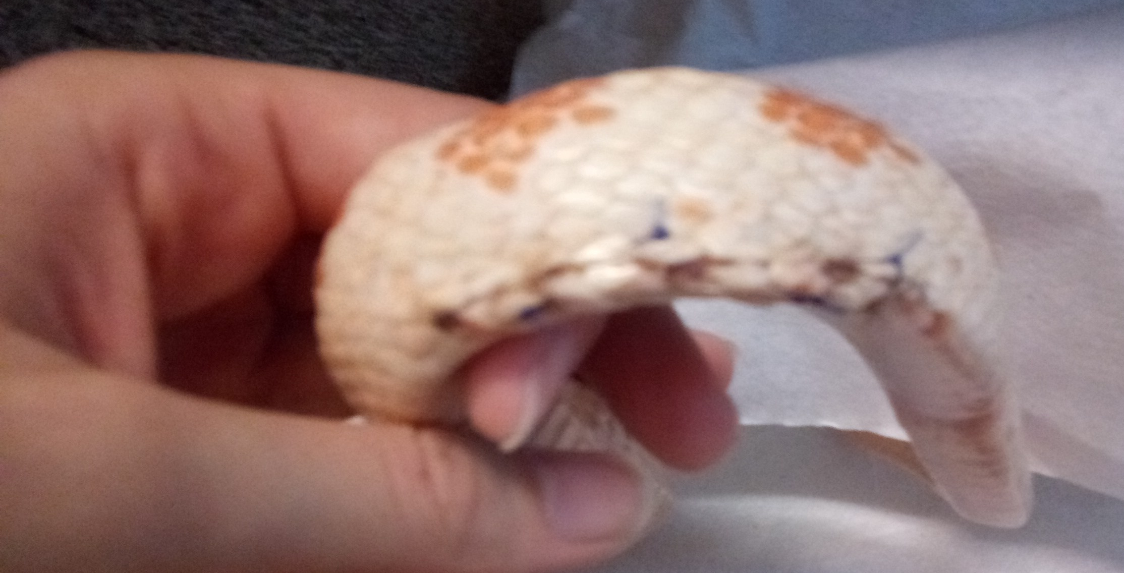
Image III: This is a blurry photo (my apologies) of William’s stitches 5 days after surgery. The blue parts are the stitches. You can see where the scales pucker; the stitches are placed in the tissue underneath this. Photo taken rather poorly by Sam Couch at WonderLab 9/23/17.
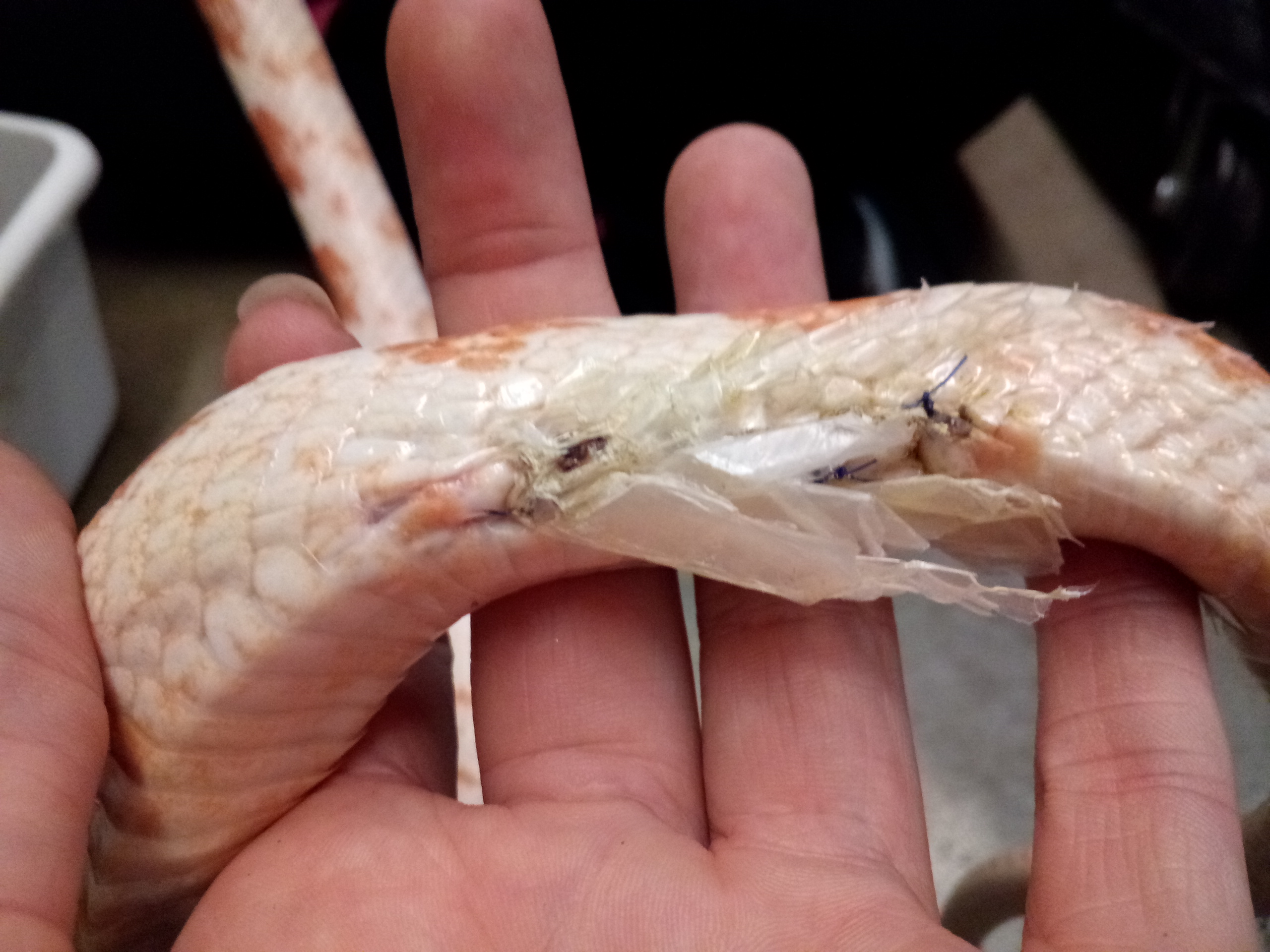
Image IV: This is Williams wound after a shed she had about a month after surgery. You can see where the stitches are holding the old skin in. We were able to remove most of this, however, the vet was got the rest out when the stitches came out. Taken by Sam Couch October 2017 at WonderLab.
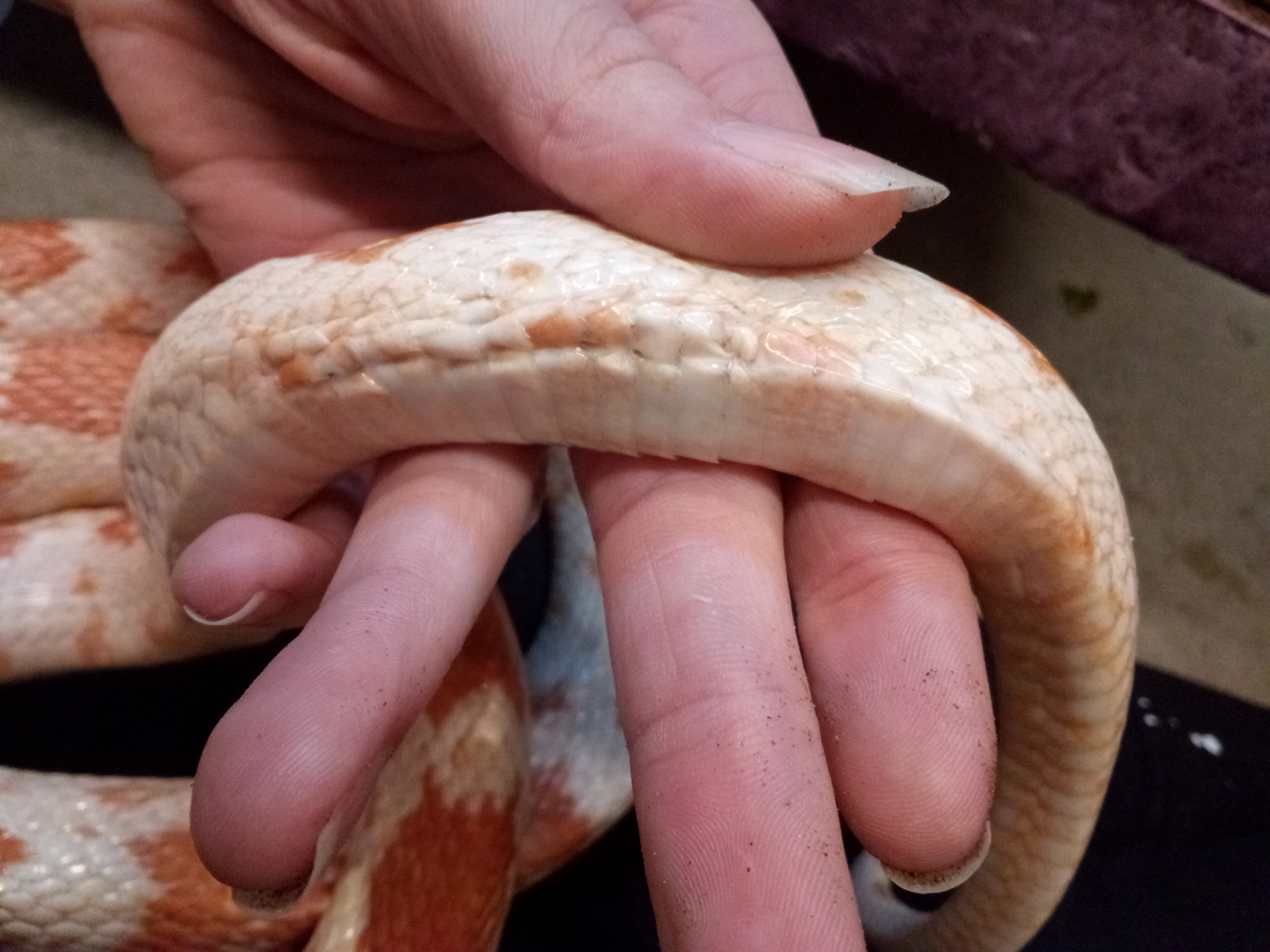
Image V: This is what William’s wound look likes today. In fact, this photo was taken 5 minutes ago by yours truly! Notice how all of the stitching is out and the scales look a bit puckered. The tissue underneath has grown back together but these scales will be a little wonky for a while and she may need some help with sheds for a few months. Taken by Sam Couch 11/17/17 at WonderLab.
While she did lose a substantial amount of weight during her ordeal, William is now eating, drinking and pooping just fine! Our team is working very hard to get her used to handling again as during post operation she was on very limited bed rest. For her safety, she will not be doing public demonstrations for the rest of the year but animal care staff works every day getting her out of her habitat and moving around again. She has been responding incredibly well to staff handling and has been getting back into the swing of things. Our goal is that she will be prepared to see all of you again in January 2018, provided her handlings go well and she gains a bit of weight back.
As is very clear here, animals receive similar medical treatments to humans. We have seen a snake get an enema, undergo a surgery that a human could have and have sutures given and removed. She went through the same tests we do like weight and height (hers is called length). While humans have grown into our own unique form, we are not so different from our scaly, slimy, furry or feathered friends. After all, we are all animals part of the same domain and we all need some TLC from time to time, right?
P.S.
For those of you familiar with William, you may be used to us called her a male. No I did not mess up the pronouns… William is in fact a female!!!! The doctors found out during surgery. No we will not change her name. She will always be our little Snakespeare.
SAM COUCH Writing to you here. I am the animal exhibits manager at WonderLab Museum of Science, Health and Technology. I found my way here from Indiana University where I got a B.S in Biology and a certificate in animal behavior with a concentration of marine science. I have done coral health assessments in the Dominican Republic and studied tropical biology in the Cayman Islands but the most important things you should know about me are that I think coral are one of the coolest animals on the planet (that we know of) but if I could be any animal in the world for a day I would want to be Cuvier’s beaked whale. In 2014 they were logged for the deepest dive, so if I were a beaked whale, I could see first hand where no human can dive!
Leave A Comment